Canine Eye Health: Understanding Cataracts in Dogs and When to Consider Surgery
Cocker Spaniels are known for their soulful, expressive eyes, but those sweet eyes are especially prone to a variety of health problems. From cataracts to glaucoma, cherry eye to chronic dryness, it’s important for every Cocker parent to understand the risks, costs, and care these conditions may require. Knowing what to watch for can help you protect your dog’s vision and comfort throughout their life.
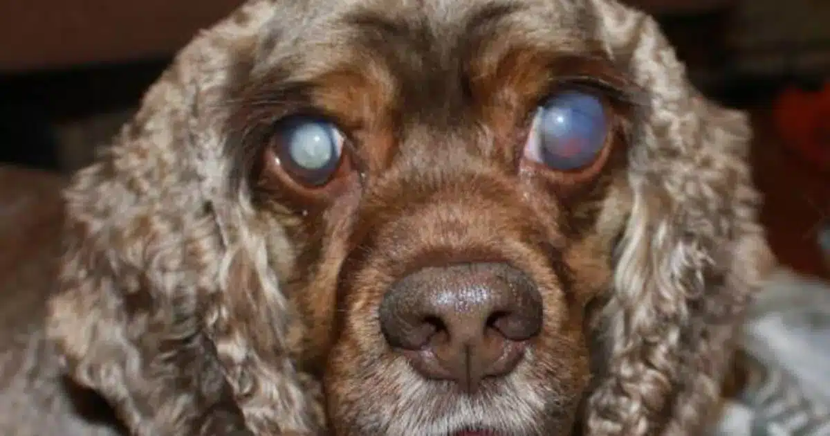
Cataracts
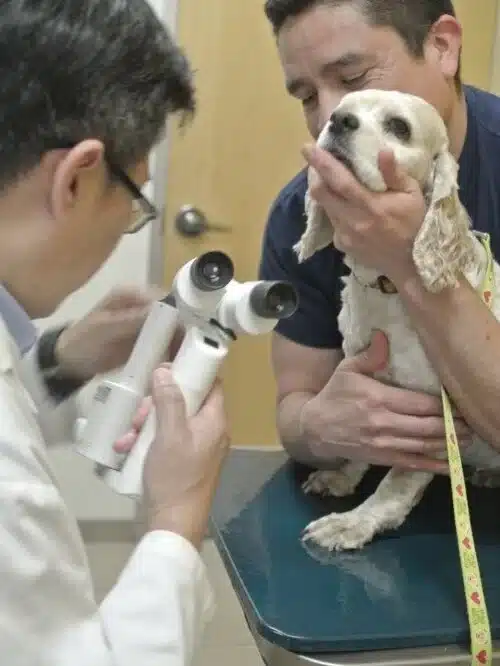
Cataracts are the opacity to the lens of the eye. The opacity can be very small (incipient cataract) and not interfere with vision. It can involve more of the lens (immature cataract) and cause blurred vision. Eventually, the entire lens can become cloudy, and all functional vision lost. This is called a mature cataract. Most cataracts in dogs are inherited, if not age-related. The cataract may develop rapidly over weeks, or slowly over years, in one or both eyes. After a lens has developed a cataract, there is no known method to make the lens clear again. Immature and mature cataracts can be treated by surgically removing them. Glaucoma occurs in 30% of all dogs who have cataract surgery. Cockers can get cataracts as young as fourteen months of age (the result of the breeder not screening out the parents for eye problems). If you are considering adopting a Cocker Spaniel, be sure you are prepared for the high expense of cataract surgery (in Los Angeles it can cost up to $3,500 per eye).
Cherry eye
Cherry Eye is the term used to refer to canine nictitans gland prolapse, a common eye condition in various dog breeds, including Cocker Spaniels, where the gland of the third eyelid known as the nictitating membrane prolapses and becomes visible. Cherry eye may be caused by a hereditary weakness in the connective tissue surrounding the gland. It is most common in puppies.
Normally the gland of the third eyelid (nictitans gland) is located behind the third eyelid in the inner corner of the eye. This gland is attached to the facial covering of the eye and eye socket by a fibrous band of tissue. Structural weakness of this attachment can lead to prolapse of the gland. The result is “cherry eye,” so called because the prolapsed gland is exposed on the surface of the eye and becomes red, inflamed and swollen.
Surgery is the usual treatment. Older methods of cherry eye correction (before the gland’s importance was known) involved simply removing the gland. Because the gland is responsible for about 30% of the eye’s tear production, the eye can eventually suffer from dryness, a condition known as “dry eye” or keratoconjunctivitis sicca, and necessitates the use of eye drops for the rest of the animal’s life. Modern methods of cherry eye correction involve repositioning of the gland to its normal location. The success rate of this type of surgery is around 80% in most breeds. Dry eye may eventually occur in 30 to 40 percent of dogs that have the gland removed, yet it may affect about 20 percent of dogs that have the gland surgically replaced. Cherry Eye “tuck” surgery (we don’t recommend the old fashioned “cut” surgery) can cost anywhere from $325 on up to $750 (these are Los Angeles area prices). There is a chance the cherry eye can pop back out again after surgery (usually in cases where the cocker is very active and isn’t kept on low activity following surgery). Ask your vet if they will offer you a reduced price if the surgery needs to be redone.
Dry Eye
Keratoconjunctivitis sicca (KCS) or “dry eye” describes the changes in the eye which result from lack of tear production.
A breakdown in the tear film and a loss of the aqueous layer causes dry eye. This loss results in dryness to areas of the corneal surface or in more advanced cases, drying to the entire corneal surface. When the cornea is deprived of oxygen and nutrients through the tear film, it rapidly undergoes destructive changes. These changes result in brown pigmentation, ulcer development, scar tissue growth, and blood vessel growth across the cornea leading to partial vision loss. Repeated eye infections and irritation to the cornea can cause severe damage. Ulceration and scarring of the cornea can eventually lead to blindness. The long-term prognosis for vision is greatly improved if the disease is diagnosed in the early stages.
A number of causes have been reported for dry eye. The majority of cases are caused by immune-mediated destruction of the tear glands. Other sources of dry eye include drug toxicity (antibiotics such as sulphadiazine and sulphasalazine can cause temporary or permanent dry eye in some animals), drug-induced reaction (atropine and topical anesthetics temporarily reduce tear production), neurological impairment (damage to the nerves leading to the lacrimal glands), removal of the third eyelid (see Cherry Eye), systemic disease (e.g. distemper), chronic conjunctivitis, trauma to the tear glands, hypothyroidism, congenital disease (some dogs are born without lacrimal glands), breed predisposition. Loss of nerve impulses to the gland due to long-standingear infections and other nerve disorders will cause a unilateral (one sided) dry eye often combined with a dry nose in some cases.
In many cases of KCS, the cause of the condition is never found. However, this does not prevent the problem from being treated.
The recognized treatment for dry eye can be the immune-suppressing drugs, Cyclosporine or Tacrolimus. This medication has provided relief of symptoms in some patients while other patients have had a marked increase in tear production.
Glaucoma
Glaucoma is defined as an elevation of intraocular pressure (IOP) that is incompatible with the health and normal function of the optic nerve. Glaucoma not only can cause complete vision loss, but also may require the need for medication or surgery. It can be painful and cause loss of the eye if uncontrolled.
Clinically,the glaucomas are divided into the three categories of primary, secondary, and congenital glaucoma. The primary glaucomas are breed-related and consist of a group of diseases characterized by an abnormal elevation in IOP due to decreased aqueous outflow without overtocular disease. These primary glaucomas are categorized further into open-angle, narrow-angle, and closed-angle glaucoma by gonioscopic examination of the iridocorneal angle, and are breed specific.
Specific therapy of patients with the glaucomas depends on the type and cause of the elevated IOP; but unfortunately most glaucomas in dogs are presentedlate in the disease when medical therapy is not very effective. Therapyis directed toward reducing intraocular pressure. This may be accomplished by either reducing the formation and secretion of aqueous humor and/or by increasing the aqueous humor outflow from the eye.
Medical therapy is usually successful for only short periods of time as the iridocorneal angle progressively narrow or closes. When primary glaucomais diagnosed and therapy initiated in one eye, prophylactic therapy should be started in the other eye to maintain the IOP within a normal range. Frequently, this consists of topical miotic therapy or beta blocker given once daily prior to darkness to prevent dilation of the pupil. One clinical study suggests that prophylactic therapy delays the onset of breed-specific glaucomas about 30 months, where as the medical control of glaucoma and preservation of vision (once the disease has been diagnosed) is only 6 months.
There are alternatives for cosmetic preservation of an eye which is otherwise blind and painful. First and foremost, it is essential to establish that vision has indeed been irreversibly lost. Secondly, the underlying cause (i.e. intraocular tumor, systemic disease leading to retinal detachment or uveitis) must be identified. Thirdly, an assessment of discomfort should be made. Usually this requires measurement of intraocular pressure and an evaluation of ocular inflammation.
- Laser Cyclophotocoagulation (CPC) involves selective destruction of a portion of the ciliary body to decrease aqueous humor production. This procedureutilizes a diode laser applied transclerally. This is the procedure of choice in cases of acute glaucoma where there is the potential to restore vision.
- Ciliary Body Ablation involves aspiration of vitreous followed by injection of a combination of gentamicin and dexamethasone. Gentamicin has been shown to be highly toxic to the ciliary body (as well as the retina) and thus results in reduced production of aqueous humor. Although this procedure is relatively inexpensive, the results are quite variable, with cataractformation and increased corneal opacity seen as well as excessive destruction of the ciliary body resulting in globe atrophy (phthisis). This procedure is generally reserved for those animals where it is necessary to avoid the depth and duration of general anesthesia required for the other surgical procedures
Click here for more information about eye problems in Cocker Spaniels.

